TERMINOLOGY OF ORAL CLINICAL AND SURGICAL PATHOLOGY
MỘT SỐ THUẬT NGỮ LÂM SÀNG VÀ MÔ BỆNH HỌC CỦA BỆNH LÝ MIỆNG
TỔN THƯƠNG SỪNG HÓA
Acanthosis: thickening of the epidermis (squamous layer); rete ridges usually extend deeper into the dermis.
--> Dày lớp gai (hoặc biểu bì), nhú biểu bì thường lấn sâu vào lớp bì bên dưới (Hình 1).
 |
| Hình 1. Nguồn: https://i0.wp.com/basicmedicalkey.com/wp-content/uploads/2017/11/A313723_1_En_2_Fig2_HTML.gif?w=960 |
Dyskeratosis: abnormal, premature keratinization of keratinocytes below granular cell layer; often have brightly eosinophilic cytoplasm.
--> Loạn sừng: sự sừng hóa sớm bất thường của các tế bào sừng/tế bào keratin nằm dưới lớp tế bào hạt, thường có tế bào chất ưa eosin màu sáng (Hình 2).
 |
| Hình 2. Loạn sừng. Nguồn: https://upload.wikimedia.org/wikipedia/commons/b/b8/Bowen_disease_%283%29.jpg |
Hyperkeratosis: thickened cornified layer, often with prominent granular layer; keratin may be abnormal; either orthokeratotic (hyperkeratosis is an exaggeration of the normal pattern of keratinization with no nuclei in the cornified layer) or parakeratotic (hyperkeratosis has retained nuclei in the cornified layer).
--> Tăng sừng: lớp sừng ở niêm mạc dày lên, lâm sàng có thể thấy niêm mạc màu trắng (như bạch sản, lichen phẳng, lưỡi lông). Thường kèm theo dày lớp hạt, lớp sừng có thể bất thường: (1) là trực sừng (orthokeratotic hyperkeratosis), (2) là cận sừng (parakeratotic hyperkeratosis) (xem các thuật ngữ tiếp theo).
 |
| Hình 3. Tăng sừng. Nguồn: https://www.ugr.es/~oncoterm/csdata/hyperkeratosisEHK-001.jpg |
Hypergranulosis: the increased thickness of the granular layer.
--> Tăng lớp hạt (Hình 1)
Orthokeratosis: thickened stratum corneum that is devoid of nuclei.
--> Á sừng/trực sừng: dày tế bào lớp sừng đã mất nhân (Hình 1). Gặp ở cả niêm mạc vốn sừng hóa hay không sừng. Có khi sử dụng lẫn lộn với thuật ngữ dày sừng (hyperkeratosis). Chính xác hơn, nên phân biệt dày sừng kiểu trực sừng (orthokeratotic hyperkeratosis) và dày sừng kiểu cận sừng (parakeratotic hyperkeratosis).
Parakeratosis: cells of cornified layer retain their nuclei, often less prominent or absent granular layer; normal for mucous membranes.
--> Cận sừng: các tế bào của lớp sừng vẫn còn nhân, thường không có lớp hạt (chỉ áp dụng đối với các biểu mô sừng hóa), đối với các niêm mạc không sừng hóa là bình thường (Hình 4).
 |
| Hình 4. Cận sừng. Nguồn: https://upload.wikimedia.org/wikipedia/commons/e/ea/Micrograph_of_early_actinic_keratosis_with_parakeratosis.jpg |
--> Teo: lớp biểu bì bị mỏng so với tình trạng bình thường (Hình 5). Khi quan sát cần đối chiếu mới phần mô lành mạnh gần đó hoặc đối chiếu với các mẫu mô trên người khoẻ mạnh.
 |
| Hình 5. Teo. Nguồn: https://ntp.niehs.nih.gov/nnl/integumentary/skin/atrophy/images/figure-001-a67792_medium.jpg |
Scale crust: parakeratotic debris, degenerating inflammatory cells, and tissue exudate on the surface of the epidermis.
--> Vảy, tróc vảy
Lichenification: thick, rough skin with prominent skin markings usually due to repeated rubbing
--> Lichen hóa: da dày, thô ráp có những vị trí nhô rõ thường do ma sát lặp lại.
Lichenoid interface change: the destruction of basal keratinocytes, causing remodeling of basement membrane zone; also bandlike lymphocytic infiltrate.
--> Thay đổi giao diện dạng lichen
TỔN THƯƠNG LỒI
Papilloma/ Papillomatosis: outward overgrowth of the epidermis with elongation of dermal papillae.
--> U nhú/ Chứng u nhú (có nhiều tổn thương u nhú): quá triển biểu bì có dạng ngón tay (Hình 6).
 |
| Hình 6a. U nhú trên lâm sàng và giải phẫu bệnh. Nguồn: https://www.intechopen.com/media/chapter/46324/media/image3.png |
 |
| Hình 6b. Mô bệnh học của u nhú. Nguồn: https://www.pathologyoutlines.com/imgau/oralcavitysquamouspapillomastojanov04.jpg |
--> Sần: tổn thương đặc, lồi, kích thước <5mm (Hình 7).
Plaque: elevated flat topped area, usually >5mm.
--> Mảng: tổn thương lồi, bề mặt phẳng, thường >5mm (Hình 7).
Nodule: solid, deeply extending lesion >5mm.
--> Hòn: tổn thương đặc, lấn sâu vào lớp mô bên dưới, kích thước >5mm (Hình 7).
 |
| Hình 7. Một số dạng tổn thương. Nguồn: https://cdn.lecturio.com/assets/Primary-skin-lesions.png |
TỔN THƯƠNG PHẲNG
Macule: circumscribed flat colored area, <1cm.
--> Dát: tổn thương phẳng có màu (khác màu biểu bì sinh lý) (Hình 7).
Patch: flat discoloration >1cm.
--> Bệt: tổn thương phẳng có màu >1cm (Hình 7).
TỔN THƯƠNG CHỨA DỊCH
Blister: vesicle or bullae.
--> Mụn/bóng nước nói chung, tổn thương chứa dịch trên bề mặt.
Bullae: fluid-filled area >5mm; either intraepidermal or subepidermal; intraepidermal bullae are due to spongiosis or acantholysis; subepidermal bullae are due to extensive papillary dermal edema.
--> Bóng nước: tổn thương chứa dịch >5mm, có thể trong lớp lớp biểu bì hoặc dưới lớp biểu bì (Hình 7, 8).
Vesicle: fluid filed area, <5 mm.
--> Mụn nước: tổn thương chứa dịch <5mm (Hình 7).
Pustule: intraepidermal or sub-epidermal vesicle or bullae filled with neutrophils.
--> Mụn mủ: mụn/bóng nước trong lớp biểu bì hoặc dưới lớp biểu bì có chứa bạch cầu (Hình 7).
 |
| Hình 8. Nguồn: https://o.quizlet.com/m.lR4eAFV6EV5PYIGlgDYQ.png |
Cyst: encapsulated cavity or sac lined by true epithelium.
--> Nang: tổn thương chứa dịch có lớp biểu mô lót toàn bộ bên trong (lòng nang) (Hình 9).
 |
| Hình 9. Nang. Nguồn: https://i.pinimg.com/originals/49/6e/cb/496ecbadef68c80ba192c665badef113.jpg |
Acantholysis: loss of intercellular connections (desmosomes) between keratinocytes; occurs in pemphigus vulgaris and related disorders; causes change in cell shape from polygonal to round.
--> (Chứng) bong lớp gai: mất các liên kết liên bào (desmosome) giữa các tế bào sừng, thường gặp trong bệnh pemphigus vulgaris (Hình 8b).
Epidermolysis: alteration of granular layer with perinuclear clear spaces, swollen and irregular keratohyalin granules, increased thickness of granular layer; different from acantholysis.
--> (Chứng) bong biểu bì (Hình 8c).
Spongiosis: intraepidermal edema, causing splaying apart of keratinocytes in stratum spinosum (resembling a sponge), vesicles due to shearing of desmosomes
--> (Chứng) phù nề lớp Malpighi (bao gồm lớp đáy và lớp bóng) (Hình 10).
 |
| Hình 10. Phù nề lớp Malpighi. Nguồn: https://media.springernature.com/lw785/springer-static/image/chp%3A10.1007%2F978-3-319-44824-4_1/MediaObjects/324456_1_En_1_Fig5_HTML.jpg |
TỔN THƯƠNG LÕM
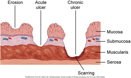
Erosion: discontinuity of mucosa causing partial loss of epidermis.
--> Chợt: tổn thương mất tính liên tục của niêm mạc do mất một phần biểu bì (Hình 11).
Ulceration: discontinuity of mucosa causing complete loss of epidermis and possible loss of dermis.
--> Loét: tổn thương mất tính liên tục của niêm mạc do mất toàn bộ lớp biểu bì và có thể một phần lớp bì (Hình 11).
 |
| Hình 11. Tổn thương lõm. Nguồn: https://o.quizlet.com/07G2dNQjXyLJbQI7cvguuw.png |
TÍCH TỤ TẾ BÀO MÁU Ở MÔ
Epidermotropism: atypical lymphocytes present in epidermis (seen in cutaneous T cell lymphoma).
--> Chứng hướng biểu bì: hiện diện tế bào limphô bất thường trong biểu bì (gặp trong liphôm tế bào T ở da, nhiễm nấm) (Hình 12).
Exocytosis: normal appearing lymphocytes in epidermis (spongiotic dermatitis).
--> Hiện tượng xuất bào: hiện diện tế bào limphô bình thường trong biểu bì (viêm da lớp bóng) (Hình 13).
: are pinpoint, round spots that appear on the skin/mucosa as a result of bleeding.--> Đốm xuất huyết: điểm tròn nhỏ xuất hiện trên da hoặc niêm mạc do xuất huyết, đường kính 1-3mm.
Purpura: extravasation of red blood cells into the skin or mucous membranes.
--> Ban xuất huyết: hồng cầu thoát mạch ở da hoặc niêm mạc, đường kính 3-10mm.
Ecchymosis
--> Bầm máu: tổn thương có đường kính >1cm. Lưu ý, một số tài liệu lấy giới hạn là 2cm để phân biệt ban xuất huyết và bầm máu.
 |
| Hình 14. Xuất huyết (hồng cầu thoát mạch) trong cơ vân. Nguồn: https://ntp.niehs.nih.gov/nnl/musculoskeletal/skel_musc/hemorr/images/figure-001-a72527_medium.jpg |
KHÁC
Calcinosis: deposit of calcium.
--> Lắng đọng canxi (Hình 15)
 |
| Hình 15. Bướu sợi calxi hoá. Nguồn: https://www.pathologyoutlines.com/caseofweek/case249image5.jpg |
--> Thể Colloid hoặc thể Civatte: tế bào sừng đang chết theo lập trình thường tròn hoặc oval, nằm ngay hoặc dưới màng đáy (Hình 16).
 |
| Hình 16. Thể Colloid. Nguồn: https://www.e-ijd.org/articles/2013/58/4/images/IndianJDermatol_2013_58_4_327_113974_f2.jpg |
TÀI LIỆU THAM KHẢO
https://www.pathologyoutlines.com/topic/skinnontumorcommonterms.html


Nhận xét